
Cranial Cruciate Ligament Injury – What is it and What is a TPLO?
This week’s blog looks into one of the most common injuries we see for rehabilitation. This is the cranial cruciate ligament injury, which is commonly operated on with a surgery known as a TPLO. We are often asked what this injury means and what a TPLO is, this blog will discuss what the cranial cruciate ligament is and what a TPLO surgery entails.
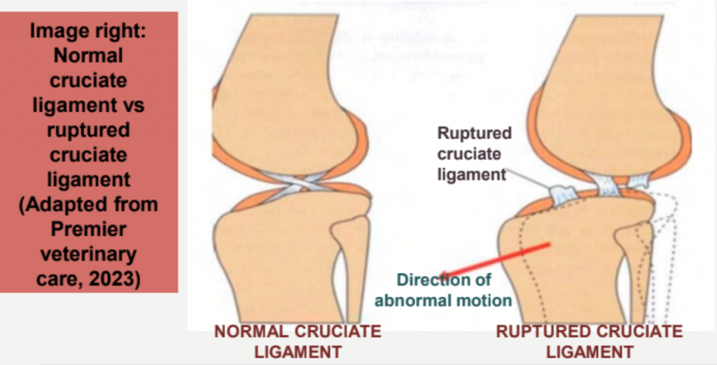
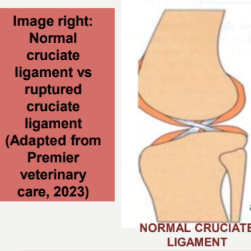
The cruciate ligaments are two bands of fibrous tissue which are found within each knee (stifle) joint. One ligament is located at the front of the knee (cranial ligament) and one at the back of the knee (caudal ligament). The ligaments join the bones above (femur) and below (tibia) to form the stifle joint. The word cruciate means to cross over. Therefore in the body, one cruciate ligaments runs from the inside to the outside of the knee joint and the other ligament runs from the outside to the inside of the knee which causes the cross over in the middle. The crossing of the ligaments helps to stabilise back to front movements of the femur against the tibia preventing one bone slipping off the other, it also prevents the two bones rotating on each other.
Dogs and cats walk on the tips of their toes rather than on the flat of their feet and so this creates a force known as the tibial thrust. This pushes the tibia forward and the femur backwards whenever they take a step. The cruciate ligaments help to neutralise the tibial thrust force however if the cranial cruciate ligament gets stretched or torn, the tibial thrust force worsens which causes the joint to become unstable. This causes pain and discomfort in our pets and the majority of the time they struggle to put weight onto the affected limb.
An option to help this problem a surgical procedure known as the Tibial Plateau Levelling Osteotomy (TPLO) is performed. This surgery aims to realign the surfaces within the knee to provide stability in the joint when the animal is walking. The femur sits on top of the tibia which is a flat, backwards leaning slope. Because the ligament has been ruptured or damaged there is risk of the femur sliding off the back of the tibial slope. The TPLO surgery involves creating a circular cut in the top of the tibia. This circular bone fragment is then rotated to cause the top of the tibia to become level, therefore ensuring the femur will no longer slip off the back of the bone. The bone fragment is fixed into position using metal bone plates, and these stay in the dog’s body for life and a new stable joint is formed.
We hope this blog has helped give you a background into the anatomy of the stifle joint to help understand what is happening and how a TPLO surgery may be an option. Over the upcoming weeks in future blogs, we shall be delving into pre and post rehabilitation for this injury, what to expect, conservative management for cruciate rupture (non-surgical management), how to prepare for the TPLO surgery and answering some of the most common questions we face as Veterinary Physiotherapists when it comes to your dogs recovery.